Computerized analysis of subcellular architecture as a tool for disease diagnosis
Abstract
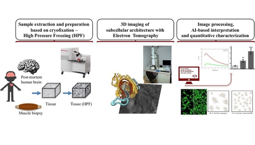
The general objective of this project is to enable the subcellular architecture (constituted by the subcellular compartments, their morphology, their spatial distribution and the relationships amongst them) as an image-based biomarker for its practical application to disease diagnosis, staging, prognosis and evaluation of the effects of pharmacological treatments. The main imaging techniques to explore subcellular architecture are electron microscopy and electron tomography (similar to computerized axial tomography -CAT- commonly used in medicine, but working at nanometric scale). They are combined with advanced protocols for tissue sample preparation that are based on cryo-fixation to preserve the native structural integrity.
This project intends to take advantage of disruptive digital technologies (artificial intelligence, advanced image processing) to develop novel computational methods for automated analysis and quantitative characterization of the subcellular architecture visualized by electron microscopy and electron tomography and the alterations associated to diseases. The ultimate aim is to enable objective and quantitative interpretation and to increase the throughput, thereby paving the way for the practical applicability of ultrastructural studies as biomarkers
Project Details
Project code:TED2021-132020B-I00
Duration:01/12/2022- 30/11/2024
Funding: 172.500 €
PI: José Jesús Fernández
Funding Science and Innovation Ministry


